07-Sep-2026
When asked about auto-immune conditions, most people would probably be able to name a few, for example Lupus or Rheumatoid Arthritis. Simply put, auto-immune conditions involve the immune system incorrectly recognising the body’s own cells as foreign, and attacking them. Less known are allo-immune conditions, where disease is caused by immune reactions to the antigens of another person, such as through a blood or organ transplant.
Fetal/neonatal alloimmune thrombocytopenia (FNAIT) is an allo-immune disease occurring in pregnancy where antibodies from the mother recognise Human Platelet Antigen-1a (HPA-1a) present in the foetus (Figure 1A). HPA-1a is part of Integrin αIIbβ3, a member of the integrin family which is key for the activation and aggregation of platelets. Recognition of HPA-1a by maternal antibodies leads to bleeding disorders in the foetus, potentially resulting in intracranial hemorrhage or perinatal death. Genetic tests that can detect mother/child incompatibility do exist, however FNAIT displays a large variation in disease severity, and severe disease cannot be predicted prenatally by current testing methods. Since treatments for FNAIT do exist, there is still a need for a diagnostic test.
Recently, scientists from different research groups across the world collaborated to try and better understand how FNAIT works at the molecular level, and why there is such variation in disease severity. The collaboration featured researchers from the Centro de Investigación del Cáncer, Leiden University, CNB-CSIC, ISBG, and Osaka University.
The group performed cryo-electron microscopy to obtain a high-resolution structure of the Fab fragment of a maternal antibody which recognises HPA-1a, in complex with integrin αIIbβ3 (Figure 1B). This antibody was originally cloned from a mother who had a fetus with FNAIT. In this case, the team first observed that the Fab fragment of maternal anti-HPA-1a antibodies reduced platelet function, rather than requiring presence of the whole antibody.
Upon further inspection, the team of researchers found that binding of the maternal Fab antibody fragment locked integrin αIIbβ3 in a closed conformation (Figure 1C-D). Previous studies have shown that extension of integrin αIIbβ3 into an open conformation is required for its function in platelet activation. Thus, this structure provides a mechanism for how anti-HPA-1a antibody binding to integrin αIIbβ3 leads to bleeding disorders.
The team then went on to investigate in exquisite detail the specific regions of the antibody fragment and integrin αIIbβ3 that interact which each other – this was made possible by solving a high-resolution cryo-EM structure. In this way, the scientists identified several key residues at the binding interface, many of which had not previously been recognised as important for this interaction. Identification of these specific contacts is important, as it opens the door for diagnostic options and therapeutic intervention. A proven treatment is with intravenous immunoglobulin which can prevent FNAIT, but it is currently not clear which women would benefit from this treatment.
Since anti-HPA-1A antibodies will be generated randomly in the mother, it is sensible to assume that different mothers will generate antibodies which bind to slightly different regions of integrin αIIbβ3, and thus inhibit its function to different degrees. Previous studies have identified that antibodies binding to other regions lock integrin αIIbβ3 in a partially extended conformation rather than fully closed as was seen here; in a partially extended conformation integrin αIIbβ3 could be expected to retain some activity. Different maternal antibodies binding to slightly different regions of integrin αIIbβ3 therefore provide a mechanism for the varying disease severity seen in FNAIT.
This mechanism has important implications for FNAIT testing. By obtaining structures of various anti-HPA-1a antibodies in complex with integrin αIIbβ3 (experimentally or through simulations), it could be possible to predict the extent to which a given antibody will inhibit integrin αIIbβ3 activity. This could then be used to develop a test to predict FNAIT disease severity using antibody samples from the mother.
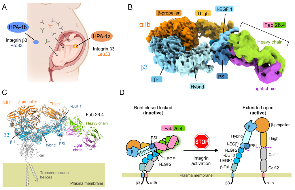
Figure 1. Structural basis of the blocking of integrin αIIbβ3 activation by the FNAIT-causing antibody 26.4. (A) When there is a mismatch between the HPA-1 antigens in the mother and the fetus, the mother may produce anti-HPA-1a allo-antibodies that cross the placenta and target fetal cells. (B) Cryo-EM map of αIIbβ3 bound to the Fab fragment of the anti-HPA-1a antibody 26.4. Regions are colored according to the different parts of the integrin and the antibody. (C) Fab 26.4 binds to the inactive conformation of αIIbβ3, as shown by the superimposition of the αIIbβ3/Fab 26.4 complex (colored) onto the structure of full length αIIbβ3 in a bent/closed conformation (PDB 8T2V, Adair et al 2023). (D) Schematic illustrating how antibody 26.4 binds to inactive αIIbβ3, blocking the activation of the integrin.
José María de Pereda commented: “Instruct-ERIC support was a great boost to this project, as this was the first time we used cryo-EM. It gave us access to state-of-the-art equipment and the expertise of facility scientists, which helped us at multiple stages of the project, from grid preparation to data collection. Processing, in particular, was not straightforward and required advanced analysis. Beyond the valuable structural data obtained, this support enabled us to establish collaborations with experts in the cryo-EM field.”
This work was made possible by access to the Cryo-EM facility and the Instruct Image Processing Center (I2PC) at Centro Nacional de Biotecnología (CNB-CSIC, Madrid, Spain), as well as the Institut de Biologie Structurale (IBS) EM facility of Instruct-FR in Grenoble, France. This access was supported by funded Instruct access proposals.
Dr Pereda concluded: “The impact extends far beyond this project: what we learned thanks to Instruct-ERIC is now allowing us to address other biomedical questions about integrins by cryo-EM and has opened up new lines of research, a clear example of how shared resources and expertise can drive scientific progress.”
The full list of authors was: José M. de Pereda, Marcos Gragera, Femke van der Meer, Francisco J. Chichón, Eleftherios Zarkadas, Ellen van der Schoot, Gestur Vidarsson, Junichi Takagi, and Coert Margadant.
Read the full paper “High-resolution cryo-EM structure of integrin αIIbβ3 bound to disease-causing maternal HPA-1a antibody that blocks integrin activation” published in Science Advances here.